Friday, 8 January 2016

Research on MERS in South Korea seems fractured...

I would be remiss if I didn't point out that the Kingdom of Saudi Arabia (KSA) is not the only country that can be seen as slow to initiate local research on local cases of Middle East respiratory syndrome (MERS), MERS-coronavirus (MERS-CoV) or the epidemiology thereof.

The outbreak in South Korea began when an incubating person flew in from Qatar, arriving 4th May. He became ill while in South Korea, with symptoms beginning on the 11th May 2015. This was to become the largest MERS-CoV outbreak outside of the Arabian Peninsula and it is still to be investigated as the overarching single event that it really was. Scientific papers, letters, media and case reports all look at small groups of specimens, this or that specific hospital, a particular slice in time or an interesting group of cases and they reach a range of conclusions. But there appears to be no over-arching all-encompassing study of the outbreak. We did eventually see such a report from the KSA in its description of four dozen early MERS cases - 13 months after the first case became ill.[4] South Korea only had 185 cases in total (exporting one to southern China) so this should not be a great task.

A recent article in The Korea Times noted that "while the initial tests were conducted on a limited number of patients, research is underway to screen clinical specimens from 32 people that will help shed better light on the matter."[1] Well, perhaps a part of the matter. [1]
The article focusses on Spike gene mutations that were found in the MERS-CoV variants from 8 human cases - which is not a remarkable discovery and has been happening since we first saw sequenced MERS-CoV from humans (see "Tracking MERS-CoV through time: a spikey problem" [2]). 

The article also goes on to say that "There is a need to focus the country’s research capability on finding the reason for the change." Those will be dollars wasted since one cannot "find the reason" for a virus mutating because that is akin to asking why we exist. 

Scientific studies can determine that the variants have mutated at certain points and they can seek out whether those changes have any effect on the efficiency of viral replication, transmission or on our immune response to infection. This will be achieved by comparison to other non-mutated MERS-CoV variant viruses and will need more than one older variant to be included so as to encompass all of the different Spike gene mutations that have been determined to date. But these studies cannot find the reason for these mutations. Yes, that is a pedantic point.
We are about 6 months out from when the final MERS case first became ill in the South Korean outbreak. This media report sadly highlights that the world is still far away from seeing comparative experimental data defining whether or not there was anything special about the MERS-CoV variants in the outbreak. Strangely though, it has already been concluded that the rapid spread of MERS-CoV in South Korea was not due to viral changes but instead because of the poor control of transmission in healthcare settings.[3] That makes this report and its message to the public both a little stale and perhaps even unnecessarily alarming.

Clearly, the KSA was not alone in needing to improve its collaboration, communication and organization of research in response to the appearance of a new virus. 

References...

  1. http://www.koreatimesus.com/mutation-detected-in-s-korean-mers-virus-govt/
  2. http://virologydownunder.blogspot.com.au/2013/08/tracking-mers-cov-through-time-spikey.html
  3. http://www.who.int/csr/disease/coronavirus_infections/situation-assessment/update-15-06-2015/en/
  4. http://www.ncbi.nlm.nih.gov/pubmed/23891402


Thursday, 7 January 2016

Zika virus disease (ZVD): 14 cases until 2007 then Yap island, Micronesia...

Update #1: 17JAN2016
Between the discovery of Zika virus (ZIKV) in 1947 and today's review subject, an outbreak on Yap Island,[1] there had only been 14 cases of Zika virus disease (ZVD) described. The keyword there is detected, or actively sought.

Some opinions on ZIKV...

In my opinion - and as the following study supports for this outbreak it describes - the confirmed ZIKV cases we heard of during 2014 are likely the tip of a vastly bigger iceberg of human infections. Many of the countries from which we have recent reports of "first" autochthonous (local) ZIKV transmission may well have had this virus in their midst for much (much, much, much?) longer since it often causes mild illness. Unless carefully looked for, ZVD is clinically very similar to other mosquito-borne virus infections. 

I hope that on the list of things to do to understand ZIKV - probably topped by investigating the suspected link between ZIKV infection and microcephaly) - is go back to stored human sera and look for traces of ZIKV or antibodies against ZIKV in these newly announced transmission zones. This will give us an idea of how long this virus has been around.

Yap Island ZVD outbreak in 2007...

In April and May of 2007, an outbreak of rash, conjunctivitis, subjective (not measured) fever, arthralgia, and arthritis occurred on an island group (the 4 Yap Islands; population ~7,400 in 2000) that comprise Yap State in the Federated States of Micronesia. Three initial patients tested positive for IgM antibodies that suggested recent dengue virus (DENV) infection (although IgM can be non-specifically triggered as a result of another infection). However. the doctors believed the symptoms were not due to DENV infection.

Samples were sent away to the United States Centers for Disease Control and Prevention and ZIKV RNA was detected in the sera of 10 of 71 (14.1%) symptomatic people using a specific reverse transcription polymerase chain reaction (RT-PCR) assay. Other viruses were considered but could not be detected using specific assays. These viruses included dengue, chikungunya, o’nyongnyong, Ross River, Barmah Forest, and Sindbis virus. The finding implicated ZIKV as the likely cause of the outbreak, and ZVD as the disease.

The result triggered a bigger investigation during which 185 suspected cases (including 49 confirmed and 59 probable) were further investigated. 66% of the confirmed and probable casess were female and these 108 people had a median age of 36y. The earliest infections occurred 15th April. Among 31 of the confirmed cases who reported symptoms, rash lasted for a median of 6d (2-14d) and arthralgia for 3.5d (1-14d). None had travelled outside of Yap.

Enzyme-linked immunosorbent assay (ELISA), an antibody detection method, was used to look for IgM antibodies indicating recent ZIKV or DENV infections. The identification of very specific infection-neutralizing antibody to these viruses was also sought and the level determined by using a plaque reduction neutralization test with a threshold value of 90% (PRNT90). Antibodies to other flaviviruses were not sought in this study.

A case definition was created:
  • A patient with suspected ZVD had acute onset of generalized macular or papular rash, arthritis, arthralgia, or nonpurulent conjunctivitis. 
    • Blood samples were requested from acute phase ( within 10d after the symptom onset) and convalescent phase (14d later)
  • A patient with probable ZVD had:
    • IgM antibody against ZIKV
    • ZIKV PRNT90 that was still detectable at a dilution of (titer) at least 1:20
    • A ratio of ZIKV PRNT90 titre to DENV PRNT90 titre was less than 4
    • Either no ZIKV RNA detected by RT-PCR or a sample which was inadequate for the performance of RT-PCR
  • A patient with confirmed Zika virus disease had:
      • ZIKV RNA detected in their serum or 
      • IgM antibody against ZIKV (detected by ELISA) and 
      • A ZIKV PRNT90 titre of at least 20 and 
      • A ratio of ZIKV PRNT90 titre to DENV PRNT90 titre of at least 4. 
173 of 200 randomly selected households were surveyed yielding 557 blood samples of which 414 (74%) had IgM anti-ZIKV antibody detected and 156 of these recalled a relevant recent illness. Among those without any IgM detected, 27 also recalled a relevant illness. Extrapolation of these figures resulted in the authors estimating that 73% of residents were infected during this outbreak with 18% of those infected having a clinical illness likely due to ZIKV infection.

An examination of 1,366 containers holding water, frequently (43%) found that they also contained 9 species of mosquito larvae/pupae and such containers were present in 87% of households. [A simple intervention method is to tip these out and ensure they cannot refill with water]. Three other mosquito species were identified to as adult mosquitoes. Aedes hensilli was the most frequently identified mosquito species overall. 

No mosquitoes tested contained detectable infectious ZIKV (examined by cell culture methods) or ZIKV RNA (RT-PCR).

So, jumping forward from the 1940s to the naughties (2007), we see a major shift in the diagnostic arsenal which supported this study. It allowed us to see just how effectively a mosquito-borne virus can apparently consume an entire island community. I wonder what IgG results - generally interpreted to indicate older earlier infection by an agent - on these same sera would yield? Would they further support the author's conclusions that a viraemic human (infected individual with ZIKV in their blood) or a virus-infected mosquito had only recently arrived from somewhere else?

Could something have changed in the levels of the vector population/species resulting in a sudden surge in virus? 

The authors note that there had not been any previous disease outbreaks of this sort reported on Yap-but were isolated cases occurring earlier? Had the virus been slowly smoldering in rare transmissions from mosquito to human/other animal to mosquito etc, for a while before building up speed and manifesting as an outbreak?  Why did none of the tested mosquitoes contain traces of ZIKV virus? 

It is also worth noting that many ZVD "cases" described in more recent months may be better defined as "suspect" and not yet confirmed. But I stand to be corrected on that.

References...
  1. Zika Virus Outbreak on Yap Island, Federated States of Micronesia
    Duffy MR, Chen TH, Hancock WT, Powers AM, Kool JL, Lanciotti RS, Pretrick M, Marfel M, Holzbauer S, Dubray C, Guillaumot L, Griggs A, Bel M, Lambert AJ, Laven J, Kosoy O, Panella A, Biggerstaff BJ, Fischer M, Hayes EB.
    N Engl J Med. 2009 Jun 11;360(24):2536-43. doi: 10.1056/NEJMoa0805715.
    http://www.nejm.org/doi/full/10.1056/NEJMoa0805715

Updates...

  1. Added 18% figure to clinical illness from population extrapolations in this paper


Wednesday, 6 January 2016

Zika virus, mosquitoes and a monkey on a platform...

UPDATED: 07JAN2016 AEST
As I noted yesterday the first published description of the isolation of Zika virus was from a study set up in the Zika forest in Uganda. It was described in print by Dr George Dick and colleagues in the September 1952 issue of Transactions of the Royal Society of Tropical Medicine and Hygiene.[1]

Rhesus macaque (Macaca mulatta) [2]
In the first study, which began April 1947, six platforms were set up in the forest canopy and upon each, a caged rhesus monkey was placed. 

The first Zika virus was identified from a monkey...

This was to be a Yellow fever virus (YFV) study since Zika forest was known to harbour a lot of previously YFV infected, antibody-positive monkeys-but stuff happens on the road to completing a good plan.

By the way: placing or penning an animal in a location known to, or under suspicion of, harbouring an infectious agent - a virus in this case - is the precess of creating a sentinel animal.

On 18th April, the daily temperature recording for "Rhesus 766" had increased to 39.7'C, rising to 40'C the next day. It was taken to the lab at Entebbe, a blood sample collected and then the primate was observed for the next month. The only sign or symptom of illness in 766 was the fever.

The blood was injected intraperitoneally (no signs of illness) or intracerebrally (became ill from day 10 post-injection) into mice and underneath the skin of Rhesus 771 (which did not develop any signs of illness).

A filterable "agent" (filters were used to remove everything but very small viruses) was recovered from the brains of the ill mice and also from the serum of 766 - it was later named Zika virus (766 strain). The virus's growth could be hampered by antibodies which developed in the serum of 766 a month later, identifying the that the Zika virus was capable of triggering a specific immune response, despite a mild illness. The more lab-savvy of you may have realised by now, that this all happened before cell culture was being used. The only way to "grow" or amplify more virus was to infect anew a susceptible host animal-which could also reveal whether the agent was capable of causing disease, so long as the experiments were suitably controlled.

A second Zika virus was found among mosquitoes...

A second virus (later called strain E/1) was acquired during a different study, but also set up for YFV. The E/1 was identified from a ground up, unfiltered preparation of Aedes africanus mosquitoes in saline which had been caught 11-12th January 1948 using a variation of the platform system. . The preparation was injected intracerebrally into 6 mice and subcutaneously into Rhesus 758 via 9 inoculations, across 2 weeks. 

Diagnosis of Zika virus by infection of animals...

Primate 758 showed no signs of illness, but the mice had - at the 7th day - so at day 8, 9 and 10 after 758's injection, blood was collected and serum injected intracerebrally into groups of 6 new mice. 
  • From the first primate sample (day 8), 2 injected mice died and virus could be passed on to additional mice from filtered preparations of their infected brain tissue
  • From the second sample (day 9), 1 mouse died (and its brain tissue filtrate could also produce new infections in mice). A second mouse was paralysed but another virus, called Theiler's mouse encephalitis virus [TMEV], a picornavirus, was identified upon further infections 
  • No mice died from the 3rd 758 inoculation of serum
  • The serum from 758 was shown to block infection of animals by Zika virus after it was first preincubated with preparations of:
    • the agent isolated from 758 serum after inoculation with the A. africanus preparation or
    • the E/1 Zika virus strain or
    • the 766 Zika virus strain
The authors concluded Zika virus was a new virus and that it triggered a specific antibody response which did not cross-react with YFV, dengue virus or the TMEV found in the paralysed mouse. 

It was also notable that disease was mild or went unnoticed in primates. Primates are usually considered to be close animal substitutes for humans in studies of disease progression. 

No studies of pregnant primates were conducted which does raise the question of whether we should consider a check-list of things to research for each and every virus capable of replicating in us.

References...
  1. Zika virus. I. Isolations and serological specificity.
    DICK GW, KITCHEN SF, HADDOW AJ.
    Trans R Soc Trop Med Hyg. 1952 Sep;46(5):509-20.
    http://www.ncbi.nlm.nih.gov/pubmed/12995440.
  2. https://www.flickr.com/photos/wild_speedy/4185543087/
Updates..
  1. Added some links and worked out some typos and layout issues.

Tuesday, 5 January 2016

Zika virus briefly...

Update #1. 05JAN2016 2030 AEST
Update #2. 30JAN2016 1430 AEST
Zika (pronounced [zÄ“k′ É™; 1] or zeek-a) virus has been in the news in recent months as it has seemingly spread very quickly from country to country, seemingly at the same time as a rise in cases of an otherwise rare human disease, microcephaly. 

The virus was first grown in the laboratory from samples from a naturally infected sentinel rhesus macaque (monkey) which were placed in cages on variously elevated platforms in the Zika ("overgrown") forest in Uganda in 1947.[18] The virus was not descried in the literature until 1952 although many others from the discoveries in this region were.[18]

I'm new to Zika virus and the study of its spread and disease so I'm on a mission to read up on it. I like to start from the beginning thus I have some of the earliest papers and will gradually read them and share with you any summaries I write up. For now, a general overview of some key bits.

Zika virus causes Zika virus disease (ZVD) or Zika fever - it's that virus versus disease thing that we have for almost all infectious diseases. Zika virus is often abbreviated to ZIKV.

From ViralZone's (www.expasy.org/viralzone,
SIB Swiss Institute of Bioinformatics) excellent
Flaviviridae page at 


ZIKV is a mosquito-borne virus that has an RNA genome (positive sense) and is enveloped by a lipid membrane (with exposed viral bits embedded-see adjacent image) surrounding an icosahedral capsid. Inside the capsid is the genome. This is the same basic structure as that found among other viruses of the family Flaviviridae of which ZIKV is a member; it falls into the genus Flavivirus of the family Flaviviridae to me a bit more precise.

ZIKV replicates in the infected host cell's cytoplasm and first makes a single protein (a polyprotein) which is subsequently cleaved up into different functional peptides.[5,8]


Aedes aegypti mosquito. One of the genus Aedes of 
mosquitoes found to host ZIKV. Other mosquitoes 

Image from CDC via Wikipedia.[9]
Some flaviviruses are borne to us (and other animals) by other arthropods - ticks apart from mosquitoes - infecting us via a virus-laden puncture/bite/injection during which virus is introduced. These viruses are all lumped together under the umbrella term of arthropod-borne viruses or arboviruses

Arboviruses replicate (grow) within the cells of the particular arthropod host, where they are amplified to higher viral loads; for example cells lining the mosquito gut in the case of Dengue virus.[10,11] Some human and animal hosts, as far as we know, cannot amplify the virus enough for it to be sucked back out by and infect the next arthropod that might come along and feed on us - these are called incidental or "dead-end" hosts.[4]

Some flaviviruses have not yet been linked to an insect host. Wikipedia maintains a great long list of flaviviruses, arthropod hosts and their mammalian and avian incidental hosts, as well as those viruses not yet linked to a human or other animal hosts.[4]

Other flaviviruses (members of that genus) you may have heard of include Yellow fever virus (YFV; the prototype virus and from where "flavi", which derives from flavus, meaning yellow/blond/golden in Latin, comes from), Dengue virus (DENV), Japanese encephalitis virus (JEV), Tick-borne encephalitis virus (TBEV), West Nile virus (WNV), St. louis encephalitis virus and Murray valley encephalitis virus (MVEV). But there are 53 species listed in the genus by the International Committee on Taxonomy of Viruses (ICTV) as of 2014 - many of which you won't have heard anything about.[3]

Generally, ZIKV causes a relatively mild illness in a proportion of those infected.[13] Signs and symptoms can include fever, rash, joint (arthralgia) and muscle (myalgia) pain, conjunctivitis, headache and jaundice but with its recent rapid spread - or more rigorous detection - a link is being investigated to a parallel rise in cases of a rare disease, detected at birth or thereafter, called microcephaly. This has been reported in some countries with ZIKV cases, but not from all. At least to date that has been the case - it may change with the new attention this disease and this virus have now attracted. 
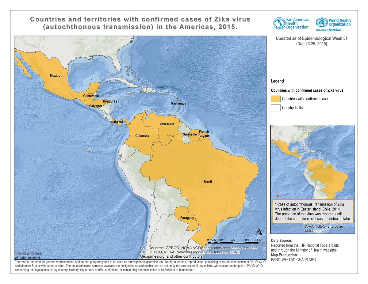
Countries and territories with autochthonous transmission.
Epidemiological Week 51 - 2015. PAHO & WHO.[23]

Microcephaly manifests, as the name suggests, as markedly smaller than normal head size and is linked to reduced brain growth in utero or brain development after birth.[6] While a link between microcephaly and viruses is not new, a link (statistical, supported with data) has yet to be found to ZIKV infection.[7] That is not to say a link will not be found, but it is awaiting the required studies. Three instances report ZIKV in amniotic fluid or in newborn tissues and one case of sexual transmission tentatively described.[19,21] Thousands of ZVD cases have reportedly been accruing on a weekly basis in Colombia alone which found its first local (autochthonous) transmission October 2015.[20] The first autochthonous reports of ZIKV infection in the Americas were confirmed in February 2014 on Easter Island, Chile.[22] In May 2015, Brazil reported discovery of its first autochthonous cases and November 2015 saw the first autochthonous circulation reports by El Salvador, Guatemala, Mexico, Paraguay, Suriname, and Venezuela.[22]

Laboratory confirmation of a suspected or probable case can be by detection of ZIKV RNA using RT-PCR in samples from an acutely infected case and by the finding of IgM antibodies 5 or more days after illness onset.[12,13] ZIKV antibody studies must be considered alongside studies of other flaviviruses which may cross-react or non-specifically flare up during infection by another related, or unrelated, virus.[14,15,16,17] The time during which virus remains in the blood may only be 3 to 5 days.[13]

But other infections can look just like ZVD including:[12]

  • dengue viruses
  • leptospirosis
  • malaria
  • rickettsia
  • group A streptococcus
  • rubella virus
  • measles virus
  • parvovirus
  • enterovirus
  • adenovirus
  • Chikungunya virus
  • Mayaro virus
  • Ross River virus
  • Barmah Forest virus
  • O’nyong-nyong
  • Sindbis viruses
So you can see that a lot of work, time and money is required for a lab asked to confirm the disease, rather than a specific viral infection.

No specific antiviral or vaccine exists for ZIKV infection - or most other viral infections.

References...
  1. http://wwwnc.cdc.gov/eid/article/20/6/et-2006_article
  2. http://viralzone.expasy.org/all_by_species/43.html
  3. http://www.ictvonline.org/taxonomyHistory.asp?taxnode_id=20141996&taxa_name=Flavivirus
  4. https://en.wikipedia.org/wiki/Flavivirus
  5. http://web.stanford.edu/group/virus/flavi/2008/flavi.html
  6. http://www.mayoclinic.org/diseases-conditions/microcephaly/basics/definition/con-20034823
  7. http://www.childrenshospital.org/conditions-and-treatments/conditions/microcephaly/symptoms-and-causes
  8. http://viralzone.expasy.org/all_by_protein/24.html
  9. https://commons.wikimedia.org/wiki/File:Aedes_aegypti_CDC-Gathany.jpg
  10. http://www.sciencedirect.com/science/article/pii/S0042682207006642
  11. http://journals.plos.org/plosntds/article?id=10.1371/journal.pntd.0001385
  12. http://www.cdc.gov/zika/hc-providers/clinicalevaluation.html
  13. http://ecdc.europa.eu/en/healthtopics/zika_virus_infection/factsheet-health-professionals/Pages/factsheet_health_professionals.aspx
  14. http://www.cdc.gov/westnile/healthcareproviders/healthcareproviders-diagnostic.html
  15. http://www.ncbi.nlm.nih.gov/pmc/articles/PMC2732478/
  16. https://books.google.com.au/books?id=73pYBAAAQBAJ&pg=PA1158&lpg=PA1158&dq=flavivirus+infection+triggers+heterologous+antibody&source=bl&ots=UYgmkBSso3&sig=CV1uAEb5NZTGfhD-hd0v6EZ7P4g&hl=en&sa=X&ved=0ahUKEwi8idCd0ZHKAhUMkZQKHXMnBNgQ6AEIOzAD#v=onepage&q=flavivirus%20infection%20triggers%20heterologous%20antibody&f=false
  17. https://books.google.com.au/books?id=BseNCgAAQBAJ&pg=PA1898&lpg=PA1898&dq=flavivirus+infection+triggers+heterologous+antibody&source=bl&ots=0JKxv695TR&sig=hvvhStq7BABzIKhzecMhFRW50cg&hl=en&sa=X&ved=0ahUKEwi8idCd0ZHKAhUMkZQKHXMnBNgQ6AEINjAC#v=onepage&q=flavivirus%20infection%20triggers%20heterologous%20antibody&f=false
  18. Zika virus. I. Isolations and serological specificity.
    DICK GW, KITCHEN SF, HADDOW AJ.
    Trans R Soc Trop Med Hyg. 1952 Sep;46(5):509-20.
    http://www.ncbi.nlm.nih.gov/pubmed/12995440
  19. http://www.forbes.com/sites/judystone/2016/01/04/zika-coming-to-america-through-mosquitoes-travel-and-sex/
  20. http://outbreaknewstoday.com/colombia-averaging-more-than-1000-zika-cases-weekly-for-the-past-month-54367/
  21. http://wwwnc.cdc.gov/eid/article/21/2/14-1363_article
  22. http://www.paho.org/Hq/index.php?option=com_docman&task=doc_download&Itemid=&gid=32405&lang=en
  23. http://www.paho.org/hq/images/stories/AD/HSD/IR/Viral_Diseases/Zika-Virus/2015-cha-autoch-human-cases-zika-virus-ew-51.jpg
Updates...
  1. Grammatical reference and additional location details
  2. Fixed some reference errors

Tuesday, 29 December 2015

Congratulations to Guinea for defeating its Ebola virus disease epidemic!

CONGRATULATIONS!!!
After a very long and painful battle with many, many losses, Guinea has stopped the Ebola virus epidemic that began there in December 2013; two years ago.[1]


From Guinea, the Makona variant of a Zaire ebolavirus spread to and throughout Liberia and Sierra Leone. For Liberia, the wait for its third declaration of freedom from any new acute human cases, must continue until 14 January.[2] Two weeks and a bit. 

Sierra Leone has remained free any human cases since early November.[3]

My family and I were out having lunch when the clock ticked over and my 10-year old boy (10M) said "Dad, no-one at any of these other tables will know how good it is to hear this news". My family has talked a lot about Ebola virus and Ebola virus disease in the past year and a half. And we've all learned a lot by talking and sharing and generally communicating. We've also been frequently reminded of all that we have. But 10M was likely very right. 

From http://virologydownunder.blogspot.com.au/2014/07/ebola-virus-disease-evd-2014-west.html
For those of us that have been watching this tragedy unfold from the sidelines since the numbers started rolling out 23 March 2014, much more so for those citizens of Guinea and Liberia and Sierra Leone who have lived and died through this, for those who went to their aid, for those who facilitated that aid and for those in countries all around the world who received and treated cases - we are happy today in a way we have not been for nearly two years. Longer for some who were involved in caring for and trying to understand and diagnose the disease in the suspected index case, a small child from Meliandou village, Guéckédou in Guinea who became ill 26 December 2013. 

While cases and clusters may yet flare up in Guinea and elsewhere in West Africa there remain many thousands of survivors who are still suffering the consequences of infection and of viral persistence.

Good luck Guinea on your 90-day period of vigilance - and beyond. You have earned some dancing!

References...

Monday, 28 December 2015

Still chatting with the demons...

It's been nine months since I took the leap out of my 23 year research career and sideways into my fantastic current role; a role which I am very lucky to have. To a researcher, luck is a very close collaborator - although one who often doesn't answer their eMail. 

Nonetheless, the demons from that former life remain to be worked through. Chocolate sultanas help at this time of year. A fantastic family is much more helpful for the other 350 or so days.

This is the first Christmas in over a decade where grant writing has not directly consumed my "holidays" (I won my first grant funding in 2004, the year after I was awarded my PhD). During the other years, the shadow of the guilt from not preparing a grant has always been in the corner of my eye. The buzz of being awarded a grant began to last for shorter and shorter periods before thoughts of the next grant barged in. These are my issues of course, and they differ for, and are coped with differently by, others

Today I notice certain research career-related media stories more often, or perhaps there are more of them, but one that I read today grabbed my attention for hitting a slew of nails on the head.[1] 

I read this while enjoying my kids playing nearby, during a humid but bird-filled afternoon - things not always noticed by me during previous Christmas seasons. The need to write, review, compile and budget filled my world view with a greater urgency than some far more important aspects of real life. I see now what a bloody fool and a complete slave to the process I had become.

Anyway, the article from today posed the question, "How do you know when it’s time to give up and move on to another career?" 

Two of a few bells that tolled for me in answer to this question quite a while ago were :
  1. Not getting national grant funding - I succeeded when NH&MRC funded about 30% of Project grant applications, but certainly not at today's 15% or less. 
  2. My publication output (Figure 1) was on a decline (ignore 2016 obviously). It was always cyclical but naturally it was strongly linked to the successes and failures of #1. I needed more, or at least better, impact factors (or whatever measure you choose to use to define "bigger" journals).
    Also, the number of citations was dropping (Figure 2) as my ageing body of work was becoming less relevant to contemporary discoveries and bigger datasets- you cannot take your foot off the gas in research 

Figure 1. Number of my publications by year. Data from Scopus.
Figure 2. Number of citations of my publications by year. Data from Scopus.
There were other reasons, but these were significant indicators to me that research was better left to those who could keep up the necessary pace of grant-getting and publishing. There are so few dollars to be had for research that they should be spent on those with the ideas to test and with the intent to achieve some long term, real benefit(s). 

The reasons for leaving research differ for everyone I imagine, but check the walls for writing. It's always on them - you may simply not be able to read the words because of the particular Kool-Aid you're being served or because of the rose-coloured glasses you've chosen to wear.

References...

  1. https://www.timeshighereducation.com/opinion/postdoc-blues-how-do-you-know-when-it-is-time-to-give-up

Friday, 25 December 2015

Avian influenza A(H7N9) virus case data in humans: more chicken scratchings

This is an example of a 2015 case announcement from the World Health Organization (WHO). I think it aims to provide information on some avian influenza A(H7N9) cases that occurred in China.

From WHO Disease Outbreak News (DON) at
http://www.who.int/csr/don/15-june-2015-avian-influenza-china/en/
I suppose it does do that in the most basic sense. Yes, if you were a casual electronic browser to the WHO disease outbreak news site (...get out more!) then you would learn of 15 additional human cases of disease presumably due to H7N9 infection. Twenty percent of these cases died and this happened within a month. 

But consider these questions for a moment:

  1. Why would you visit the WHO to learn of this, if you were not seeking some actual detail and evaluation of risk?
  2. If you were a casual browser, I expect you would come away from this with some out-of-context concerns about a bolus of cases in such a short period, spanning a wide age range and occurring across considerable geographic distance. Should you be worried? Is this the precursor to some larger outbreak? Each difficult to answer from this very small cross-section of information.
  3. These public data are relied upon by some when they write papers or release infectious disease reports - so why not include key - yet deidentified - demographic detail in a line list format - remember MERS-CoV in South Korea anyone? That WHO list [4] was messy [5] but it was a step forward for those outside the WHO network who wanted free, publicly available basic data, quickly
In my opinion, the premier emerging disease tracking and publishing - at least in terms of accessible, basic, rapid, searchable, freely accessible and up-to-date emerging infectious disease information - is the team at FluTrackers. Keep your predictive modelling - I'd trade it all in for a clone army of these guys any day! The FluTrackers line list on H7N9 includes some of these cases[6]...and as you can see in the snippet below, even their scouring of the media from China does not help to fill in the data gaps on these cases...
From FluTrackers' H7N9 line list at
https://flutrackers.com/forum/forum/china-h7n9-outbreak-tracking/143874-flutrackers-2013-15-human-case-list-of-provincial-ministry-of-health-government-confirmed-influenza-a-h7n9-cases-with-links?t=202713 [6]
So why does the WHO bother with this information at all? I can't speak for them. But one reason may be because it is relied upon by those who study and prepare for the emergence of new or re-emergence of old infectious agents.[1] These people consume this sort of information to track what's happening outside their own back yard and to weigh the risks that a new bug may come knocking at the gate thanks to a speedy international plane flight. But when the host country is slow or perhaps even reticent to identify key case details, a knowledge gap emerges and may widen. This particular gap has been growing since 2014's H7N9 3rd wave. Perhaps since the 2nd wave.

When sufficient - or any - detail is lacking, then it comes down to the public to look for any answers they seek...by themselves. In this case, that has been FluTrackers & Co; this source has proven itself very worthy for this and for other viral threat monitoring, but cannot be expected to fill this need indefinitely.[2,3]

H7N9 is a good example of an absence of obvious change to the flow of information that the world's public, citizen scientists and its more professional scientists receive about new infectious threats. And this is all a bit strange because I was sure we'd heard a lot about the need to do much, much better on this sort of tracking and chatting in 2014/5 during one of the biggest modern moments of being "caught with our pants down" - the Ebola virus disease epidemic.

Time and viruses wait for no person. Be faster.

Ho. Ho. Ho.

References...

  1. http://ecdc.europa.eu/en/publications/Publications/RRA-Influenza-A-H7N9-update-four.pdf
  2. http://www.ncbi.nlm.nih.gov/pubmed/24885692
  3. http://www.ncbi.nlm.nih.gov/pmc/articles/PMC4140362/pdf/jphr-2012-2-e29.pdf
  4. http://www.who.int/emergencies/mers-cov/MERS-CoV-cases-rok.pdf?ua=1
  5. http://virologydownunder.blogspot.com.au/2015/06/matching-mers-case-identification.html
  6. https://flutrackers.com/forum/forum/china-h7n9-outbreak-tracking/143874-flutrackers-2013-15-human-case-list-of-provincial-ministry-of-health-government-confirmed-influenza-a-h7n9-cases-with-links?t=202713

Thursday, 24 December 2015

Seasons Greetings from VDU...

Wishing you all a very Happy Christmas and a safe, happy and healthy New Year.


Tuesday, 15 December 2015

One man has a lot of MERS on his mind....

Professor Christian Drosten heads a sizable, very friendly and diverse team of scientists seeking to understand all aspects of the Middle East respiratory syndrome coronavirus (MERS-CoV). 

And not just understand, but also to detect, educate, vaccinate and generally interrupt MERS-CoV transmission in camels and to and between humans. 

That's a heckuva lot to think about.

From the First WHO-EMRO Training Workshop on MERS-CoV Laboratory Diagnostics in cooperation with the Central Veterinary Research Laboratory (CVRL). 

Sunday, 13 December 2015

Where I am this week...

With my personal thanks to the University Hospital Bonn's organizational team, the World Health Organization - Eastern Mediterranean Regional Office, the Central Veterinary Research Laboratory (CVRL) and the United Arab Emirates Ministry of Health.